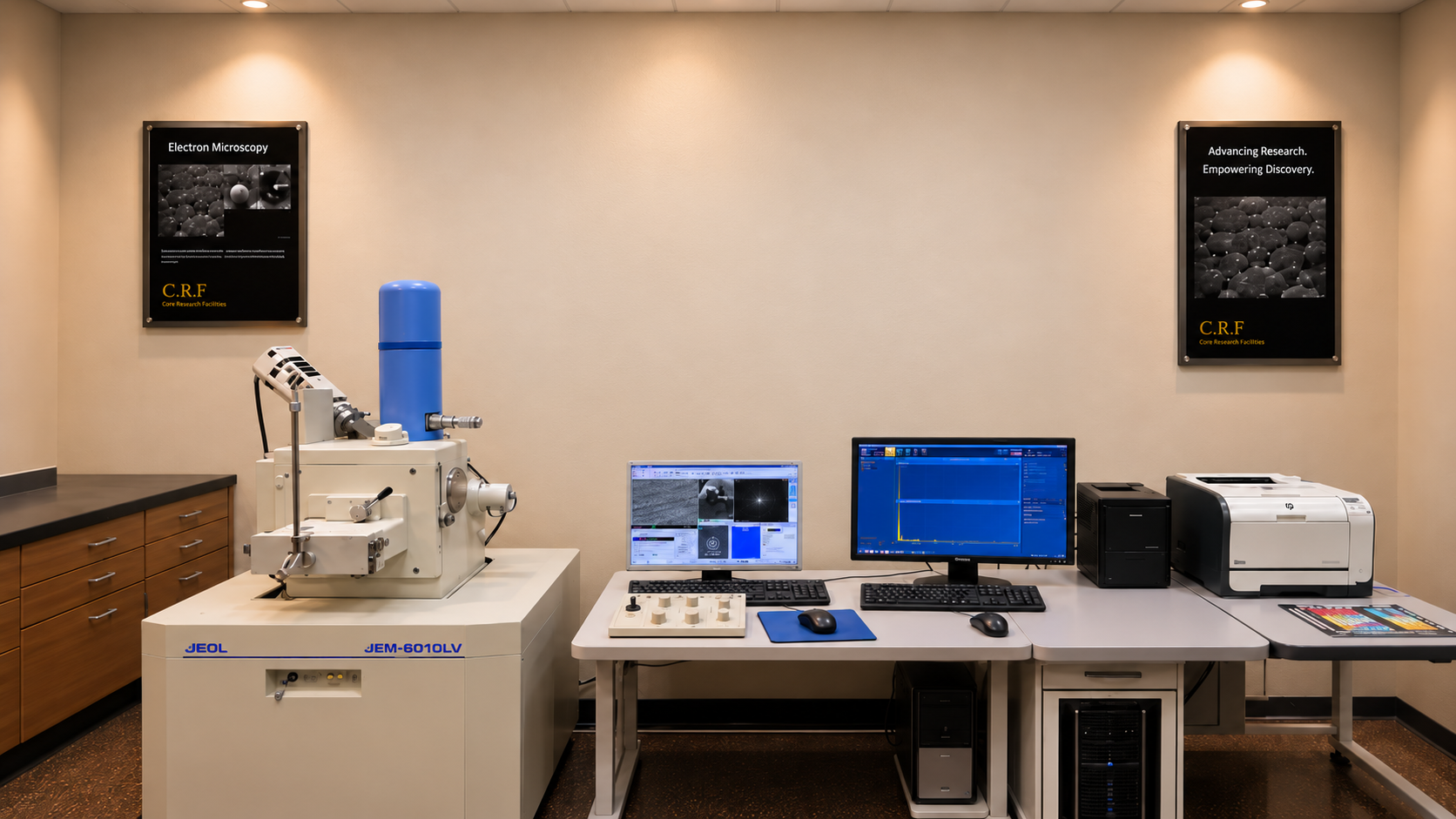
Scanning Electron Microscopy (SEM) JEOL JSM-6610LV
Scanning Electron Microscopy (SEM) is an advanced electron microscopy technique used to characterize the surface structure, morphology, and microstructural properties of materials. The JEOL JSM-6610LV operates by scanning a focused electron beam across the sample surface and detecting signals generated through electron–sample interactions. It provides high-resolution images revealing surface features, particle morphology, grain structures, fractures, and material defects. The instrument supports both high-vacuum and low-vacuum operating modes, allowing the examination of conductive and certain non-conductive specimens with reduced sample preparation. When integrated with Energy Dispersive X-ray Spectroscopy (EDS), the system enables qualitative and quantitative elemental analysis. SEM is widely used in materials science, metallurgy, geology, ceramics, nanotechnology, and industrial failure analysis.
Capabilities
High-resolution surface morphology and topography imaging
Microstructural characterization of materials
Secondary Electron (SE) and Backscattered Electron (BSE) imaging
High-vacuum and low-vacuum sample analysis
Particle size, shape, and distribution analysis
Grain structure, fracture surface, and defect examination
Qualitative and quantitative elemental analysis using EDS
Elemental mapping and compositional characterization
Applications: Metals, alloys, minerals, ceramics, polymers, coatings, composites, and corrosion products
Facilities
Model: JEOL JSM-6610LV
Manufacturer: JEOL Ltd.
Scanning Electron Microscope (SEM) with low-vacuum capability
Electron accelerating voltage range of 0.3–30 kV
Secondary Electron (SE) and Backscattered Electron (BSE) imaging capabilities
Energy Dispersive X-ray Spectroscopy (EDS) system
High-vacuum and low-vacuum operating modes
Computer-controlled image acquisition and processing
Elemental spectrum acquisition and compositional analysis
Digital imaging and microstructural characterization
Location Equipment In-charge
Core Research Facilities Name: Mr. Hatim Dafalla Mohamed Dafalla
Building 15 LAB# 2203 Phone: 860-3545
E-mail: dmhatim@kfupm.edu.sa