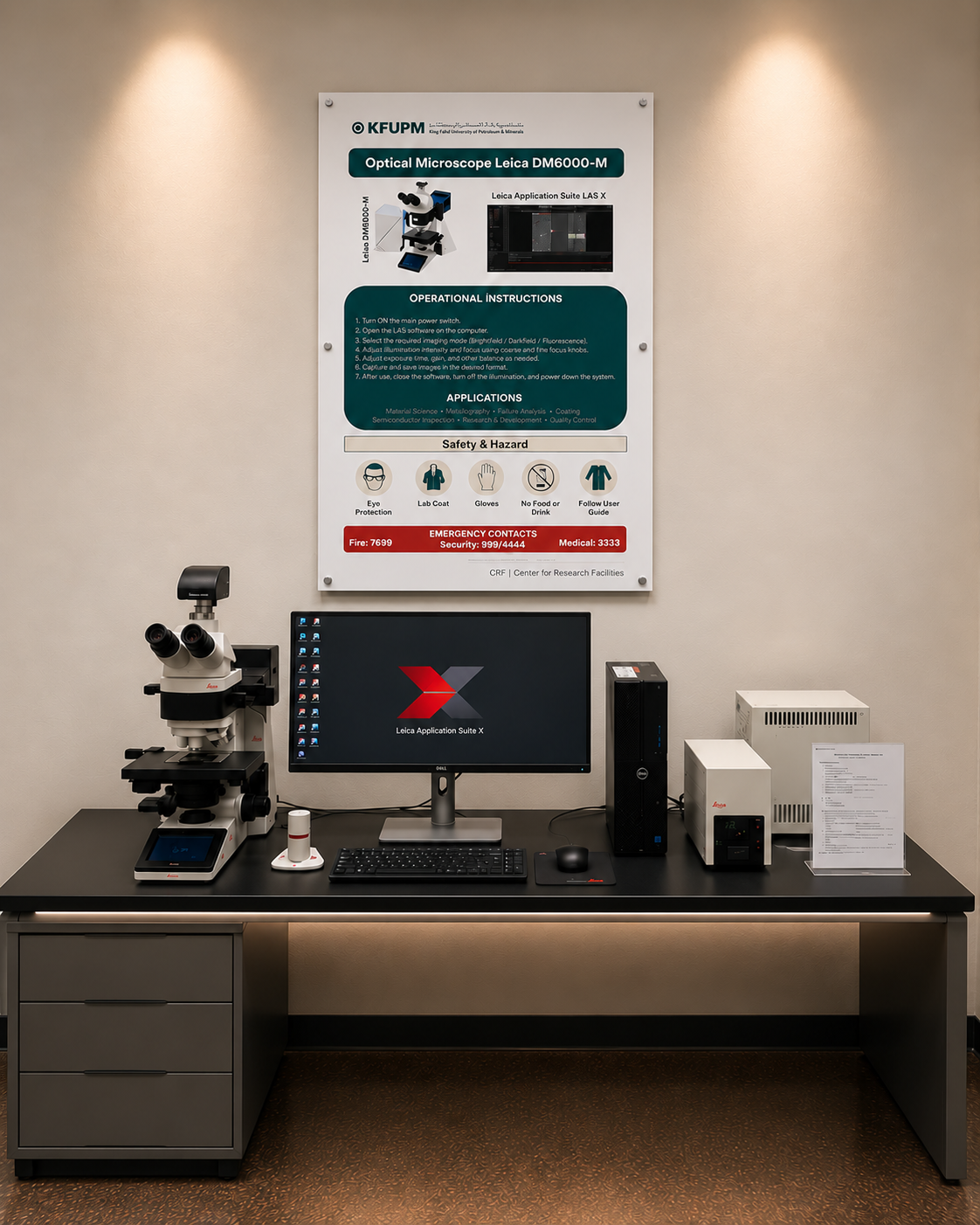
Optical Microscope
Optical Microscopy is a widely used materials characterization technique that utilizes visible light and precision optical lenses to magnify and examine the surface features and microstructural properties of materials. The Leica DM6000 M is an advanced research-grade optical microscope designed for detailed inspection and analysis of metallic, ceramic, semiconductor, and other material specimens. It supports multiple illumination and contrast techniques, including brightfield, darkfield, polarized light, and differential interference contrast (DIC), depending on the installed configuration. Integrated with Leica Application Suite X (LAS X) software, the system enables digital image acquisition, measurement, documentation, and microstructural analysis. Optical microscopy is widely used in metallography, materials science, failure analysis, semiconductor inspection, coating evaluation, and industrial quality control.
Capabilities
High-resolution optical imaging and surface inspection
Microstructural and metallographic characterization
Grain size, shape, and distribution analysis
Surface defects, cracks, and fracture examination
Coating thickness and cross-sectional analysis
Brightfield and darkfield optical imaging
Polarized light and differential interference contrast (DIC) imaging, where configured
Digital image acquisition and dimensional measurements
Applications: Metals, alloys, ceramics, polymers, coatings, semiconductors, composites, and industrial materials
Facilities
Model: Leica DM6000 M
Manufacturer: Leica Microsystems
Advanced research-grade optical microscope
Motorized microscope platform for precise imaging and positioning
High-quality optical system for detailed material examination
Reflected-light illumination for opaque material samples
Multiple optical contrast techniques, depending on configuration
Digital camera system for image acquisition and documentation
Leica Application Suite X (LAS X) imaging and analysis software
Computer-controlled imaging and measurement capabilities
Surface morphology, grain structure, and dimensional analysis
Metallographic examination and coating characterization
Location Equipment In-charge
Core Research Facilities Name: Ms. Gadeer Alonizan
Building 15 LAB# 2205 Phone: 860-5356
E-mail: gadeer.onizan@kfupm.edu.sa